Molecule Viewer
Scientists share the 3D shapes of molecules as files of atom positions: proteins and DNA in the Protein Data Bank, small molecules as SDF, MOL or XYZ files.
- Lane
- 7, Atoms
- Kind
- Built from a recipe
- Your own
- Open a structure, or fetch one by its PDB code
- Sound
- Yes
- Licenses
- CC0
How to play
Tap two atoms for their distance, a third for the angle.
Its controls
| Control | Kind | Range | Start |
|---|---|---|---|
| Measure an example | Tap | ||
| Measure | Button | ||
| Structure | List | 7 choices | Crambin (1CRN), a small plant protein |
| Draw as | List | 5 choices | Best for this structure |
| Color by | List | 6 choices | Element (CPK) |
| Hydrogens | Setting | ||
| Water | Setting |
About it
Scientists share the 3D shapes of molecules as files of atom positions: proteins and DNA in the Protein Data Bank, small molecules as SDF, MOL or XYZ files. This viewer draws every atom of such a file with splats, in the usual colors: carbon gray, oxygen red, nitrogen blue.
Pick a sample, open your own file, or type a PDB code such as 1MBN and press Fetch in the Splash tab; the entry is read only when you ask, and its title, authors and fetch time show beside it. Draw it as a cartoon, balls and sticks, space-filling atoms or a smooth surface, colored by element, chain, residue or B-factor. Tap two atoms for the distance between their centers in ångströms, a third for the angle in degrees. Positions are an experiment's best fit, not a photograph.
Details
- Your own files
- .pdb, .ent, .cif, .mmcif, .sdf, .sd, .mol, .mdl, .xyz
Is it right?
The viewer draws the atom positions a structure file gives (PDB, mmCIF, SDF/MOL or XYZ), and measures distances and angles between atom centers from those positions. The readers are checked against known values: atom counts, the same structure read from two formats, crambin's peptide bonds, backbone angles and disulfide bridges, and caffeine's C=O bonds. The cartoon, the surface and the colors are drawings of that data; where they simplify, it is said below.
Correct
Atoms are where the file puts them: the first model, the first alternate location, the same positions whether the structure comes as PDB or mmCIF.12
The PDB reader takes the fixed columns of ATOM and HETATM records; the mmCIF reader takes the _atom_site table. Crambin (1CRN) read both ways gives the same 327 atoms, elements, positions and B-factors.
Correct
Distances are in ångströms and angles in degrees, between atom centers; crambin's peptide bonds measure 1.33 Å and its N–CA–C angles 111°, as standard protein geometry says.34
A distance is the straight line between two atoms' positions; an angle is taken at the middle atom from the two directions to the others. Across crambin's 45 peptide bonds the mean C–N length is within 0.01 Å of 1.329 Å, and the mean N–CA–C angle within 1° of 111.2°.
Correct
Bonds come from the file where it lists them, and otherwise from distances with standard covalent radii.567
SDF/MOL bond blocks and PDB CONECT records are read as given. Otherwise two atoms are bonded when they are closer than the sum of their covalent radii (Cordero et al. 2008) plus 0.45 Å, and a hydrogen keeps only its nearest partner. In crambin this finds exactly its three disulfide bridges (Cys 3–40, 4–32 and 16–26), each about 2.04 Å; caffeine read as XYZ gets the same 25 bonds its SDF lists.
Correct
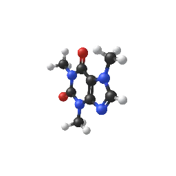
Double bonds from an SDF file are drawn as two sticks; caffeine's two C=O bonds are 1.22 Å long.89
The bond order is read from the file's bond block; caffeine's ideal coordinates from the PDB's Chemical Component Dictionary give C=O bonds of 1.216 and 1.218 Å.
Simplified
Helices and strands come from the file's records; when a file has none they are worked out from backbone hydrogen bonds, and the splash says so.10
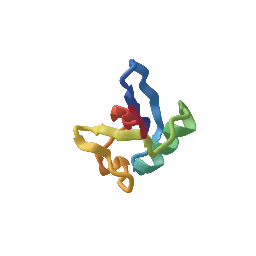
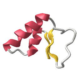
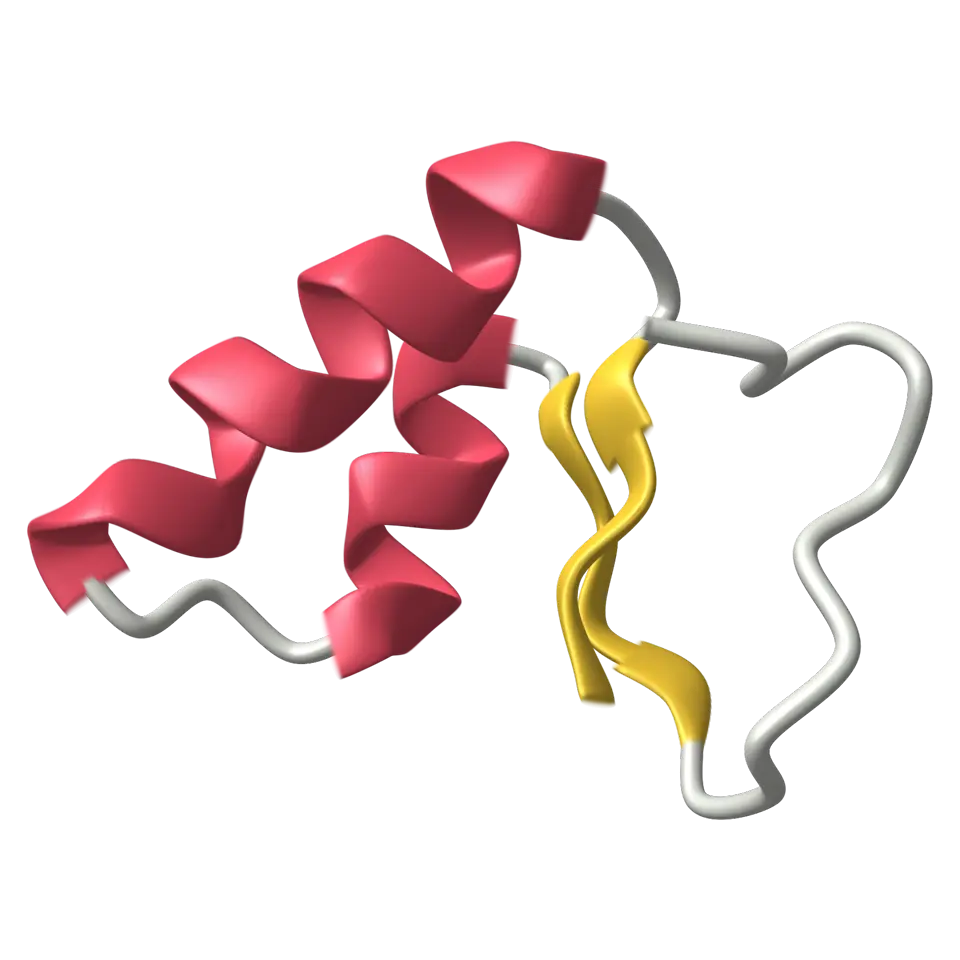
HELIX and SHEET records (PDB) or _struct_conf and _struct_sheet_range (mmCIF) mark the residues. Without them, a simplified DSSP finds backbone hydrogen bonds with Kabsch and Sander's electrostatic energy (below -0.5 kcal/mol) and builds helices from turns and strands from bridges, up to 3,000 amino acids. For crambin the file gives helices 7–19 and 23–30 and strands 1–4 and 32–35; with the records removed, the worked-out version agrees on more than three quarters of the residues.
Correct
Space-filling atoms are drawn at their van der Waals radii (Bondi), in the familiar CPK colors.1112
Radii from Bondi (1964): H 1.20, C 1.70, N 1.55, O 1.52, S 1.80 Å and so on; Mantina et al. (2009) for main-group elements Bondi left out; 2.0 Å for elements with neither (the splash says so). Colors are Jmol's CPK colors, carbon a little lighter for the dark stage.
Simplified
The molecular surface is close to the surface a water molecule can touch, but smoothed.13
Each heavy atom adds exp(B(1 − d²/R²)) with R its van der Waals radius, so a lone atom's surface lies exactly at R; the surface is drawn where the sum reaches 1, sampled on a grid. Between crowded atoms the sum fills the gaps, as the solvent-excluded surface of a 1.4 Å probe does, but deep crevices come out shallower.
- 1Atomic Coordinate Entry Format Version 3.3, Coordinate Section, wwPDB
- 21CRN: Water structure of a hydrophobic protein at atomic resolution, RCSB Protein Data Bank
- 3PROCHECK Operating Manual, Appendix A: Engh & Huber parameters, CCP4 (SLAC mirror)
- 4Ångström, Wikipedia
- 5Covalent radius, Wikipedia
- 6UniProt P01542, Crambin, UniProt
- 7Disulfide, Wikipedia
- 8Carbonyl group, Wikipedia
- 9CFF, caffeine, RCSB Protein Data Bank (Chemical Component Dictionary)
- 10DSSP (algorithm), Wikipedia
- 11Van der Waals radius, Wikipedia
- 12CPK coloring, Wikipedia
- 13Accessible surface area, Wikipedia
- Simplified
- The cartoon is a drawing: a ribbon through the CA atoms (phosphates for DNA and RNA) with set widths, not a measured shape.
- Simplified
- The surface is a blobby Gaussian surface over the heavy atoms, close to the solvent-excluded surface but smoother.
- Simplified
- Big structures are drawn with fewer splats: one Gaussian per atom, and past the budget only the atoms on the outside (said in the Splash tab).
- Simplified
- Only the first model and the first alternate location of each residue are shown.
- Simplified
- A flat 2D drawing opened as MOL or SDF gets a 3D shape built by Splashery's embedder; its lengths and angles are estimates, and the splash says so.
Sources and credits
| Work | By | License |
|---|---|---|
| Crambin, snapshot of October 5, 2026 | W. A. Hendrickson and M. M. Teeter (the structure); M. M. Teeter, PNAS 81, 6014 (1984) | CC0 1.0 (wwPDB data) |
| Green fluorescent protein, snapshot of October 5, 2026 | M. Ormö and S. J. Remington (the structure); M. Ormö, A. B. Cubitt, K. Kallio, L. A. Gross, R. Y. Tsien and S. J. Remington, Science 273, 1392 (1996) | CC0 1.0 (wwPDB data) |
| Hen egg-white lysozyme, snapshot of October 5, 2026 | R. Diamond, D. C. Phillips, C. C. F. Blake and A. C. T. North (the structure); R. Diamond, J. Mol. Biol. 82, 371 (1974) | CC0 1.0 (wwPDB data) |
| B-DNA, twelve base pairs, snapshot of October 5, 2026 | H. R. Drew, R. M. Wing, T. Takano, C. Broka, S. Tanaka, K. Itakura and R. E. Dickerson (PNAS 78, 2179, 1981) | CC0 1.0 (wwPDB data) |
| Caffeine, snapshot of October 5, 2026 | the wwPDB Chemical Component Dictionary (ideal coordinates) | CC0 1.0 (wwPDB data) |